There are certain things the medical community thinks it knows: like how the bones in the wrist move and how to treat it when it’s injured. Then along come researchers such as those from Brown Medical School/Rhode Island Hospital who make science reconsider previous knowledge.
Under a five-year study funded by the National Institutes of Health, researchers at Brown Medical School/Rhode Island Hospital are using a combination of computer tomography (CT), new Geomagic reverse-engineering software, CAD/CAM solid modeling tools, and computer animations to study the human wrist in completely new ways. Early discoveries indicate that the wrist doesn’t move in previously accepted manners, and widely practiced treatments of wrist injuries could be dramatically improved.
Current knowledge of wrist kinematics and pathologies is based on clinical experience and years of research using cadavers. Cadaver-based studies possess inherent limitations, namely the inability to simulate physical stress loads on the wrists and real injuries.
Joseph Crisco, Director of the Orthopedic Bioengineering Laboratory at Brown Medical School/Rhode Island Hospital, and his colleagues are one of the first groups of researchers to gather data on wrist movement from live subjects. The process is non-invasive and uses data from CT scans of the human wrist through a full range of motion, revealing 3D images, which traditional X-rays do not.
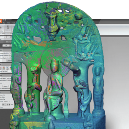
Once the scan data is collected, it is exported to Mayo Foundation’s Analyze software to extract the contours of the small bones present in the scans. Crisco and his team have developed programs that can generate point cloud data from the extracted bone contours. The point-cloud data is imported into Geomagic Wrap reverse engineering software, where the data is turned into polygonal models and NURBS surfaces.
Crisco’s group also combines scanned point cloud data of varying resolutions to create the final 3D models of the wrist bones. The surfaces of the higher-resolution scans are automatically aligned within Geomagic Wrap with corresponding point clouds from other positions and saved as new files. Using these files, the team creates accurate and easily viewed digital models of completed wrists in multiple positions.
“Without accurate digital models, we wouldn’t be able to quantitatively study various wrist positions and motions,” says Crisco.
Crisco’s team then imports the digital wrist models into SOLIDWORKS CAD/CAM software for additional fine-tuning. The final CAD data is output to a rapid prototyping machine, where physical models of the wrist are created. The models are extremely accurate, within two degrees of rotation and 0.2mm of dimension, and preserve the bone orientation of each wrist position.
Animation Reveals Complex Motion
Brown Medical researchers also use the digital models to create 3D animations of wrist movement. Proprietary mathematical algorithms help the team fill in the gaps between scanned positions by calculating and predicting the range of motion – much like a flip-book animation. The animations help the researchers see, three-dimensionally, how the relations between wrist positions play out in real life.
Based on the animations, Crisco has found that wrist motion and function are more complicated than previous theories indicate. Current descriptions of wrist motion suggest that the eight wrist bones move as three columns or two rows. Instead, Crisco has discovered that each bone has a separate pattern of motion that is associated with each unique direction of wrist motion.
“As we continue to prove our hypotheses correct,” he says, “we are generating an understanding of the increasingly complicated motion of the carpal bones. Three-dimensional animations have been the only way for us to visualize these findings.”
The Potential to Revolutionize Treatment
The Brown Medical team is studying injured wrists as well as healthy ones to determine if current diagnosis and treatment methods can be improved. They are examining soft tissue injuries, such as scapholunate ligament tears, which are often the consequence of a fall on an outstretched hand. Current evaluations of ligament tears use X-rays to examine the gap between the scaphoid and lunate bones. If the gap between is wider than expected, a ligament tear is the most probable diagnosis.